
Patient Education
Diagnosis of CM and SM
Widespread use of magnetic resonance imaging (MRI scans) has resulted in more rapid diagnosis and has also uncovered relationships between the disorders. An MRI of the spine may reveal SM, and one of the craniocervical junction may detect CM. While the disorders do not necessarily occur in tandem, a comprehensive examination may uncover CM and SM, as well as Ehlers-Danlos syndrome (EDS), scoliosis, tethered cord, spina bifida, arachnoiditis, or other related conditions.
Did you know… that approximately 350,000 Americans have CM? Many also have SM, and others have SM from other causes.
MRI
Before and After Surgery
Chiari malformation
Before surgery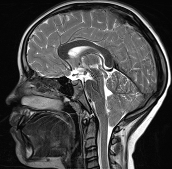
- Cerebellar tonsils are herniated through the foramen magnum
After surgery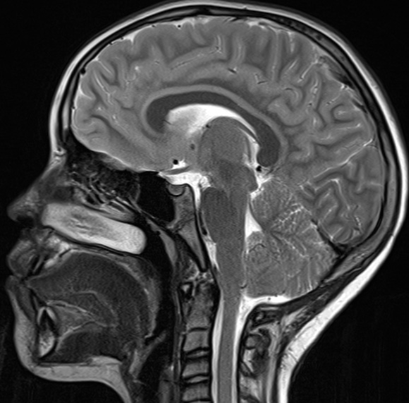
- Posterior fossa has been decompressed, allowing for more room for the tonsils
Chiari with syringomyelia
Before surgery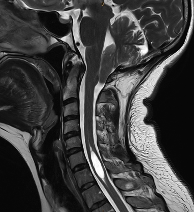
- Cerebellar tonsils have herniated into the spinal canal, impeding the flow of cerebrospinal fluid (CSF)
- Syrinx in the spinal cord
After surgery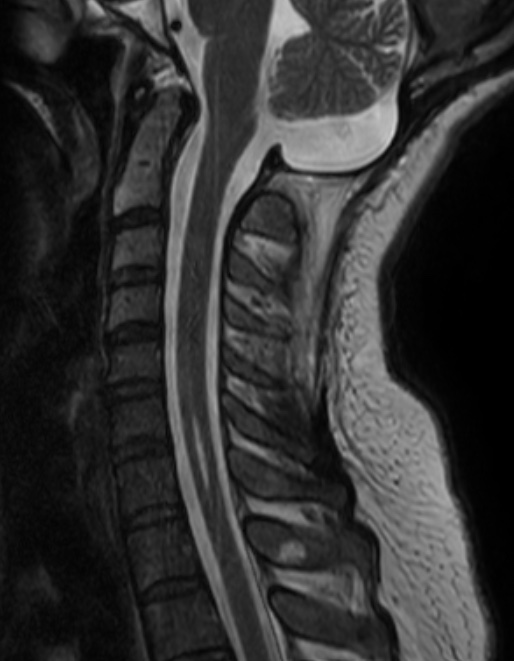
- CSF flow has been restored
- Syrinx has collapsed