Column of HopeChiari & Syringomyelia Research FoundationWorking Toward a Life Without Pain

Research Update

Summer 2025 Research Update

August 2025

Where Column of Hope's research stands, from the labs in Sydney: what we set out to solve twenty years ago, what the team has proved since, and the step in front of us now.
  1. Chiari and syringomyelia research update
Column of Hope – Summary of  Research Efforts in
Sydney, Australia
    Slide 1 — tap to see it full size

    Most of Column of Hope’s research efforts occur in two collaborative research centres in Sydney.
  2. Chiari malformation
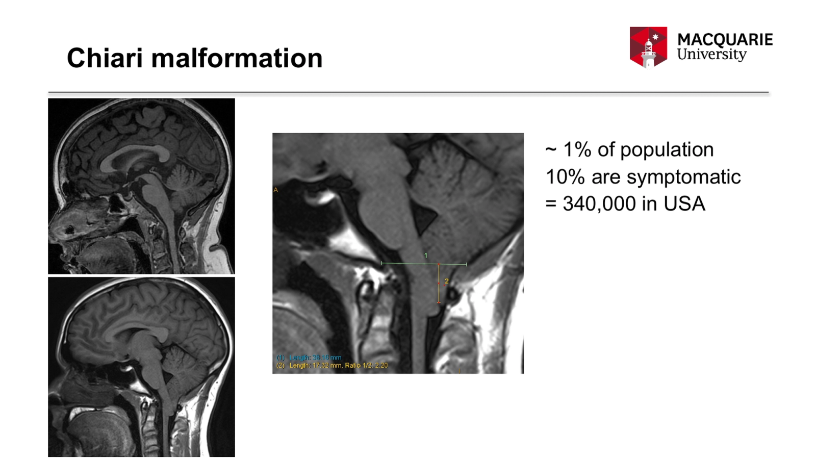
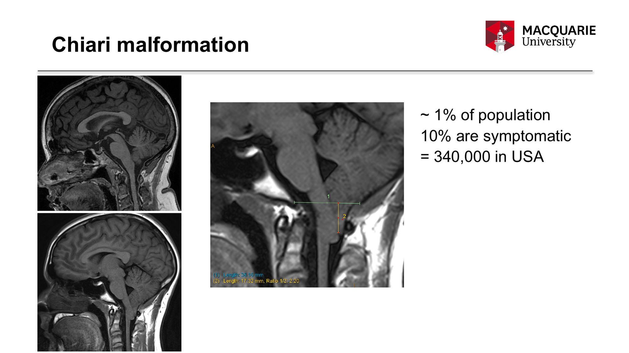
~ 1% of population
10% are symptomatic
= 340,000 in USA
    Slide 2 — tap to see it full size

    We were faced with a challenge to draw focus on solving issues, as the disorders were thought to be rare disorders. In the early 2000s, the NIH only acknowledged that there were 20,000 Americans with symptomatic Chiari malformation (CM) despite surgical counts supporting the 340,000-patient population. The image with the measurements shows how the tonsils of the brain is herniated down into the spinal column area causing the symptoms.
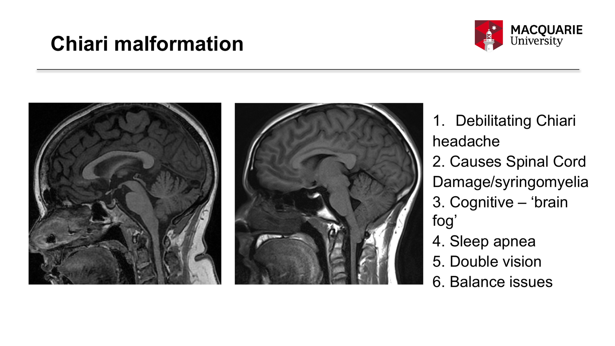
  3. Chiari malformation
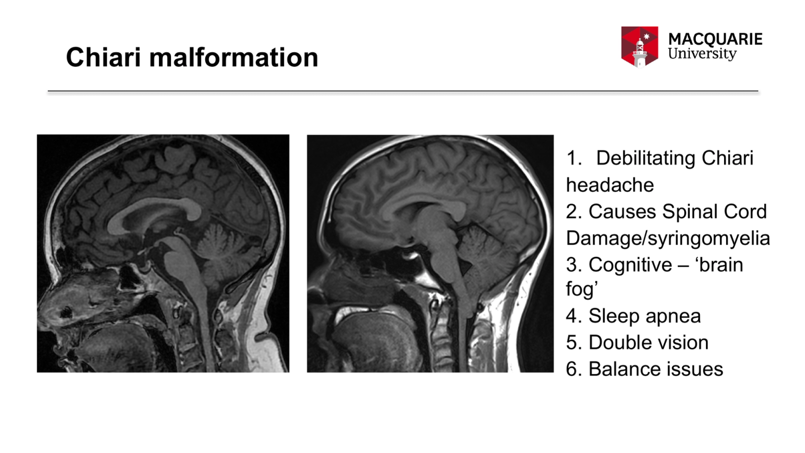
Debilitating Chiari
headache
2. Causes Spinal Cord
Damage/syringomyelia
3. Cognitive – ‘brain fog’
4. Sleep apnea
5. Double vision
6. Balance issues
    Slide 3 — tap to see it full size

    Chiari malformation alone can be debilitating, but it often causes syringomyelia with a whole new set of pain and paralyzing symptoms. The image on the right shows the brain tonsil herniation indicating Chiari malformation.
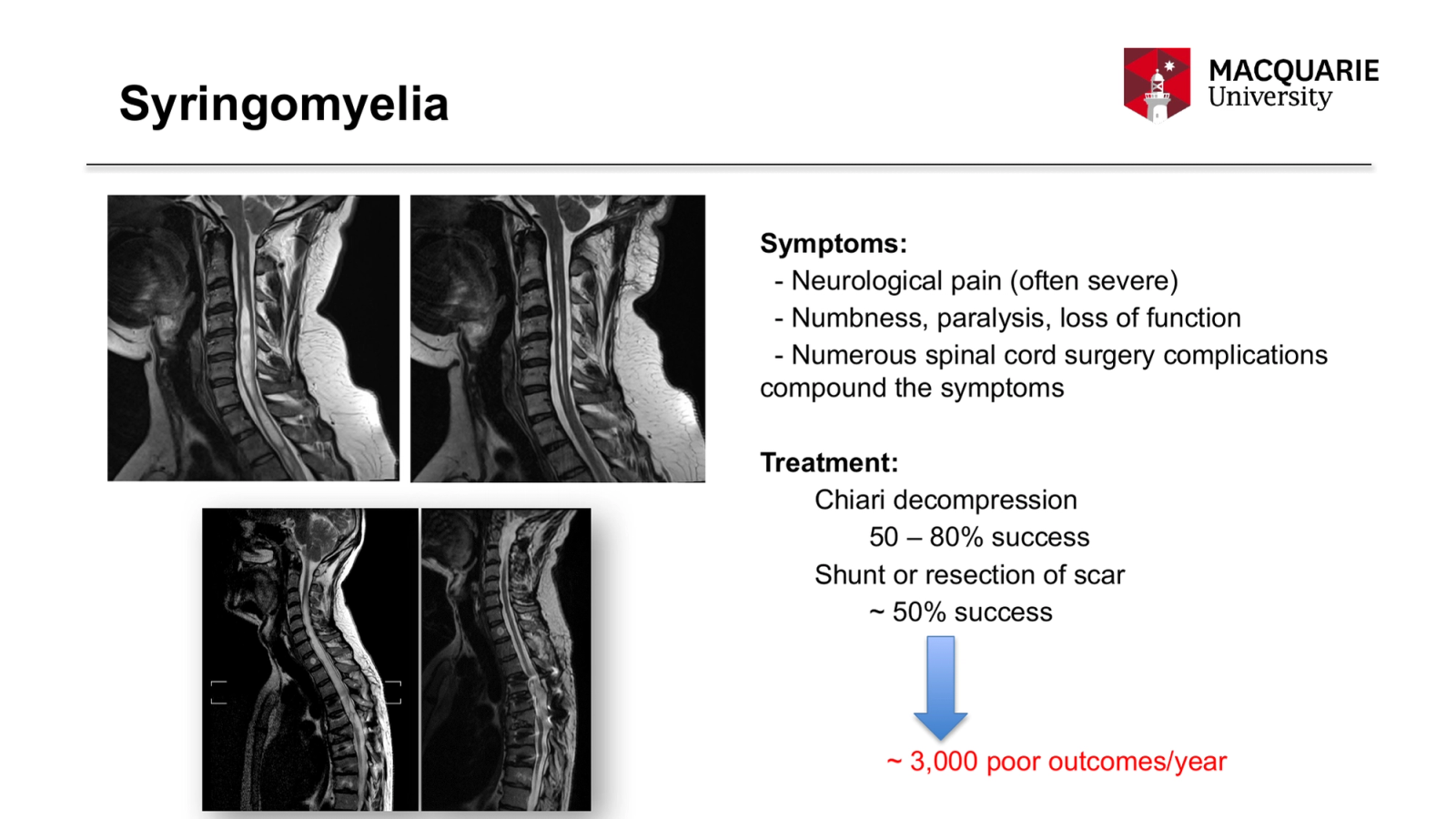
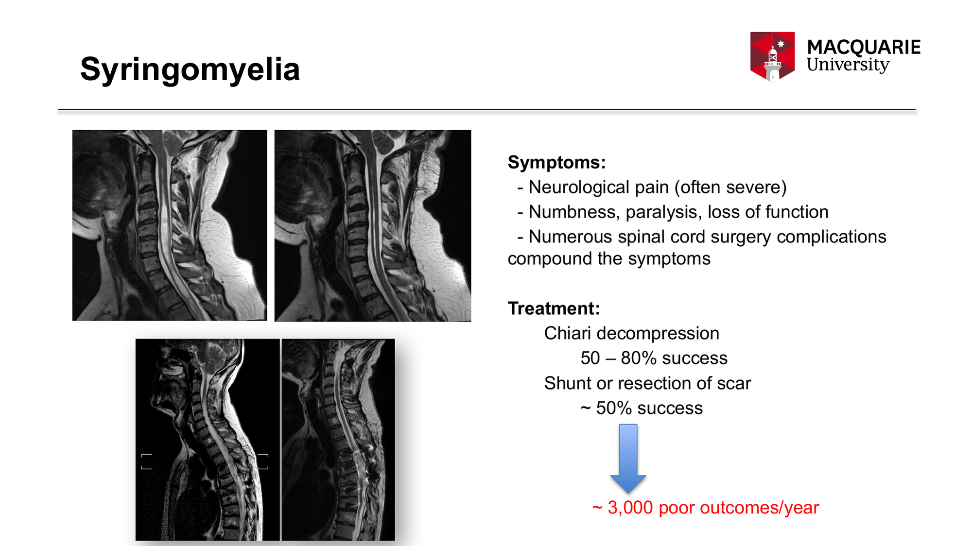
  4. Syringomyelia
Symptoms:
- Neurological pain (often severe)
- Numbness, paralysis, loss of function
- Numerous spinal cord surgery complications compound the symptoms
Treatment:
Chiari decompression
50 – 80% success
Shunt or resection of scar
~ 50% success
~ 3,000 poor outcomes/year
    Slide 4 — tap to see it full size

    It is our contention that while we agree with the 3,000 estimate of poor outcomes per year, we feel there are likely another 3,000+ of marginal outcomes. We at Column of Hope believe truly successful surgical results occur significantly less than 50% of the time.
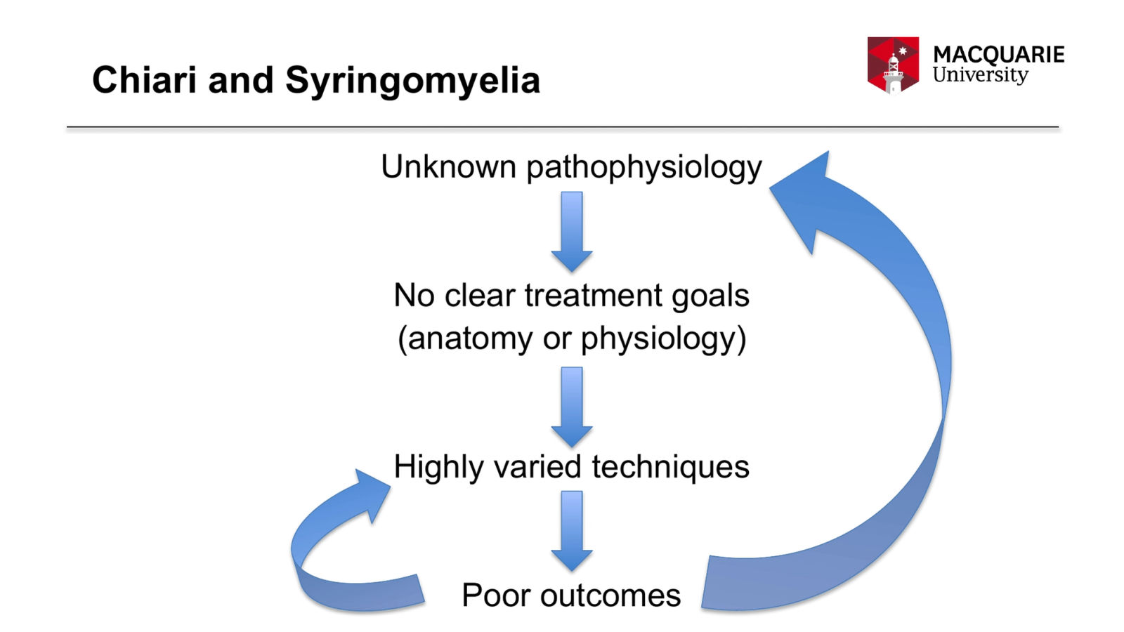
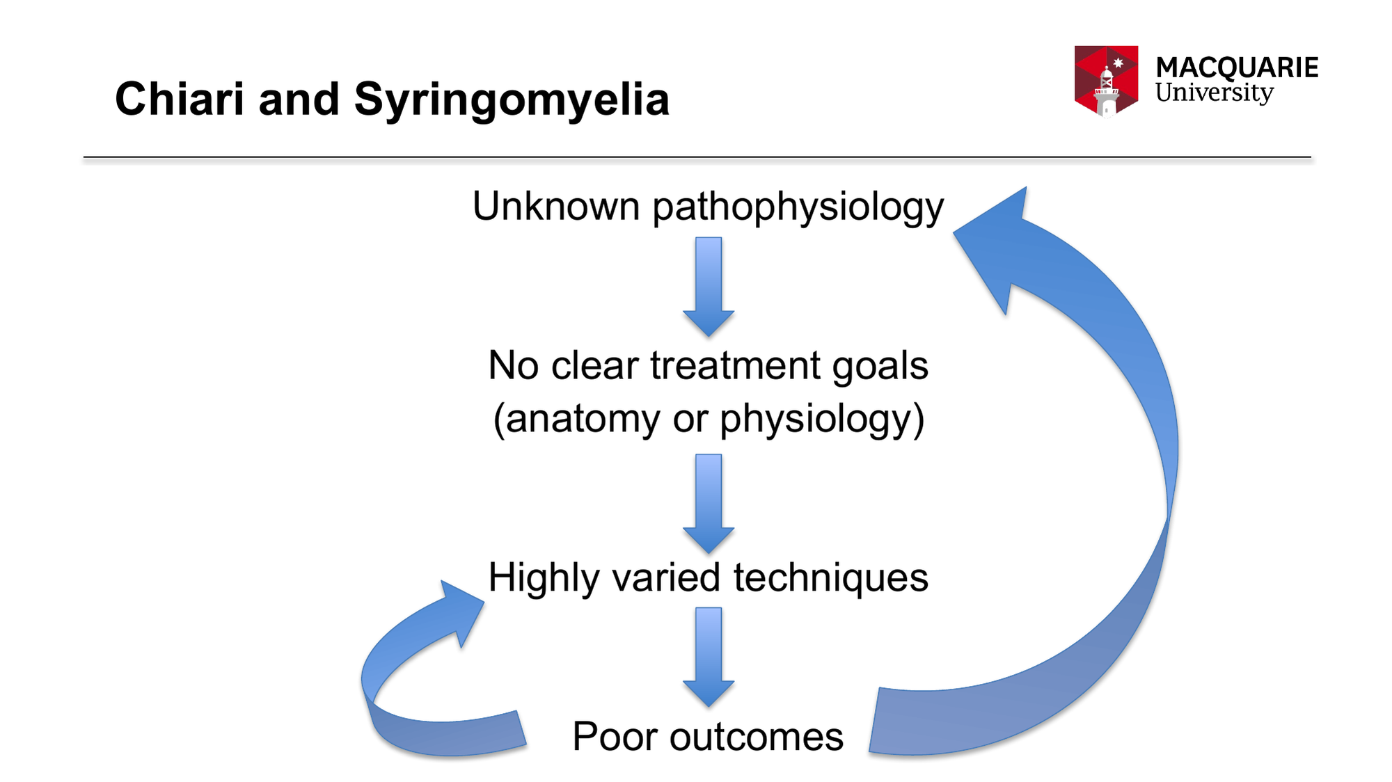
  5. Chiari and Syringomyelia
Unknown pathophysiology
No clear treatment goals
(anatomy or physiology)
Highly varied techniques
Poor outcomes
    Slide 5 — tap to see it full size

    This is where we started 20+ years ago! Root causes and the physiology of the disorders were not known. Of course, with so many unknowns, each surgeon had to wing it, especially after problems arose with the initial surgery. This process results in poor outcomes, which continues the unsatisfactory loop. Column of Hope was formed knowing the cycle had to be broken, surgeons needed to know the pathophysiology of the disorders in order to treat them, and the false assumption that the disorders were rare left research unfunded. Column of Hope borrowed techniques from the for-profit world to bootstrap its early research.
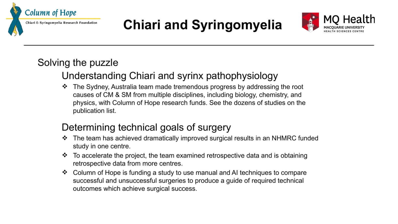
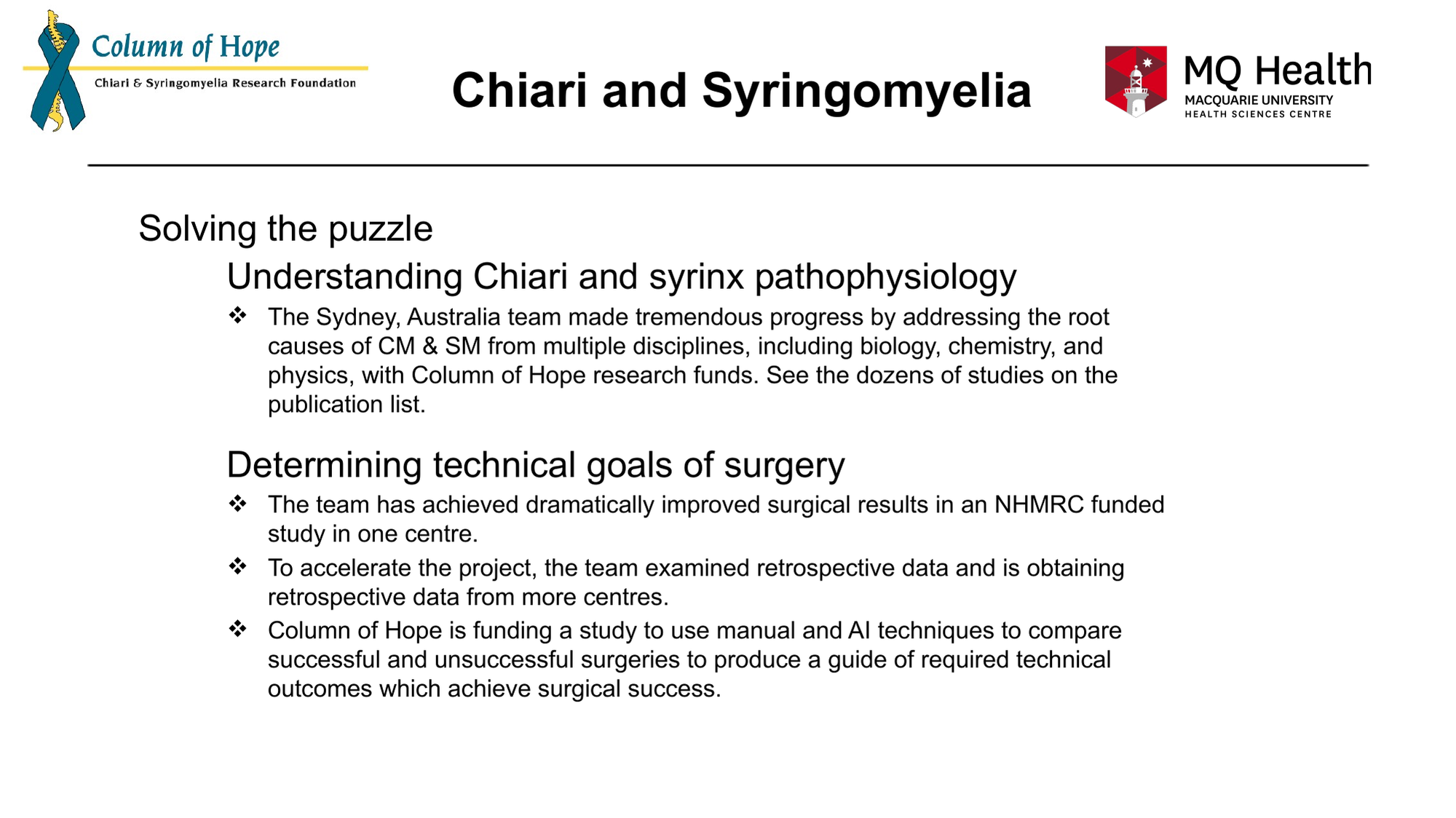
  6. Chiari and Syringomyelia
Solving the puzzle
Understanding Chiari and syrinx pathophysiology
The Sydney, Australia team made tremendous progress by addressing the root causes of CM & SM from multiple disciplines, including biology, chemistry, and physics, with Column of Hope research funds. See the dozens of studies on the publication list.
Determining technical goals of surgery
The team has achieved dramatically improved surgical results in an NHMRC funded study in one centre.
To accelerate the project, the team examined retrospective data and is obtaining retrospective data from more centres.
Column of Hope is funding a study to use manual and AI techniques to compare successful and unsuccessful surgeries to produce a guide of required technical outcomes which achieve surgical success.
    Slide 6 — tap to see it full size

    It’s a major accomplishment to achieve surgical success in one centre. It's also a major undertaking to define these technical outcomes and allow patients in all centres to achieve those outcomes.
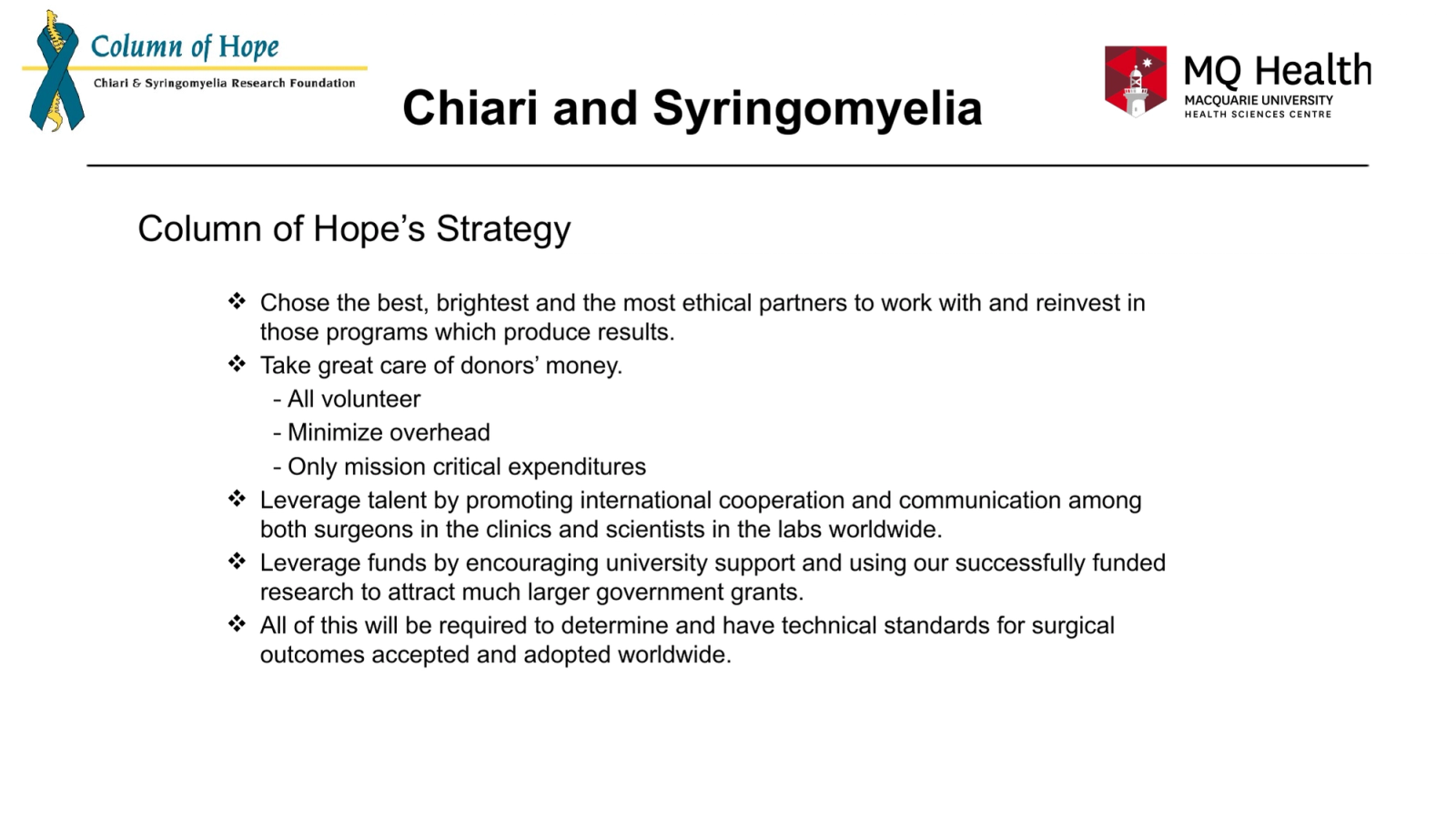
  7. Chiari and Syringomyelia
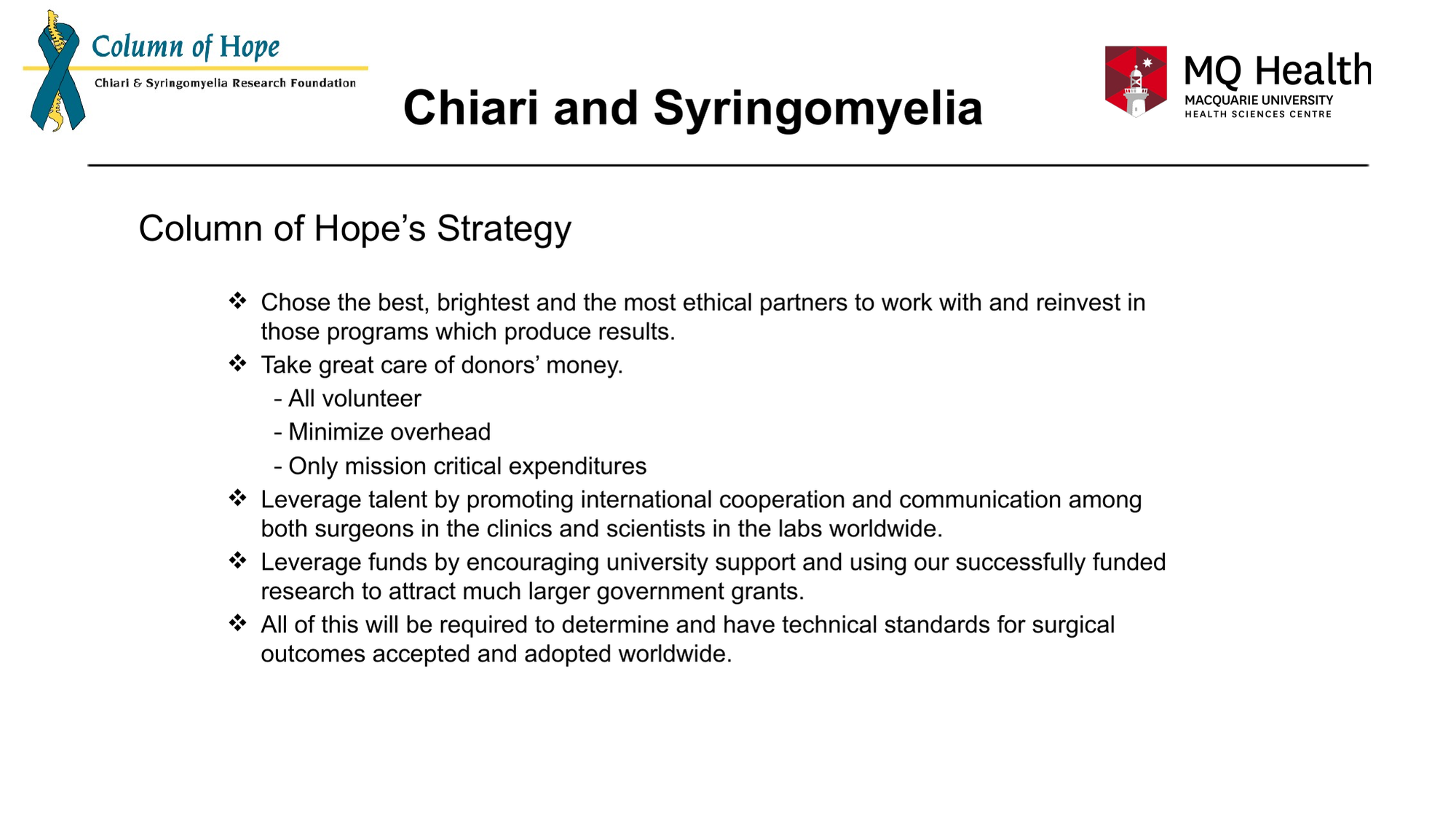
Column of Hope’s Strategy
Chose the best, brightest and the most ethical partners to work with and reinvest in those programs which produce results.
Take great care of donors’ money.
- All volunteer
- Minimize overhead
- Only mission critical expenditures
Leverage talent by promoting international cooperation and communication among both surgeons in the clinics and scientists in the labs worldwide.
Leverage funds by encouraging university support and using our successfully funded research to attract much larger government grants.
All of this will be required to determine and have technical standards for surgical outcomes accepted and adopted worldwide.
    Slide 7 — tap to see it full size

    This slide highlights our bootstrapping strategies, which are similar to those used in successful start-up biotech companies.
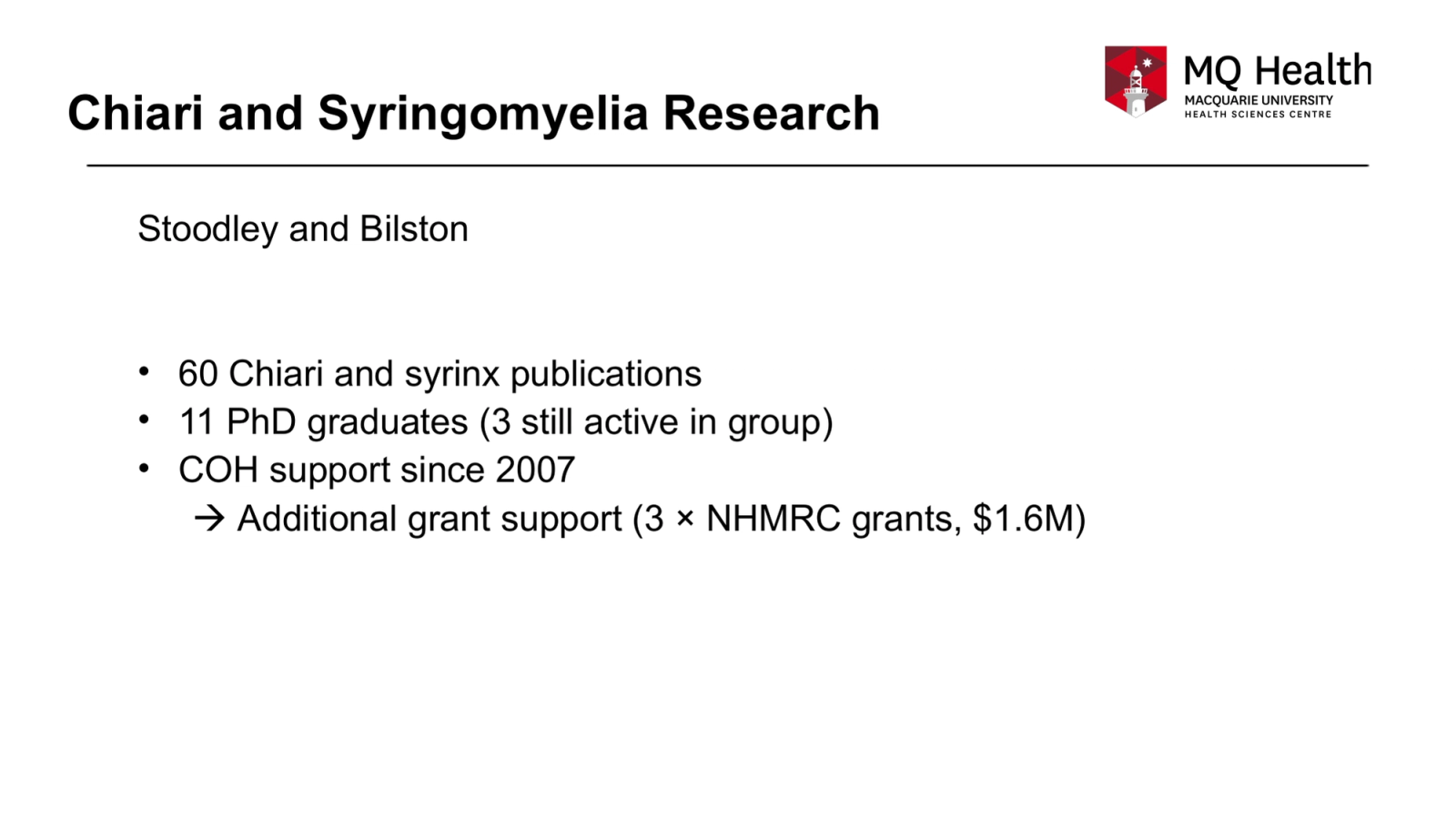
  8. Chiari and Syringomyelia Research
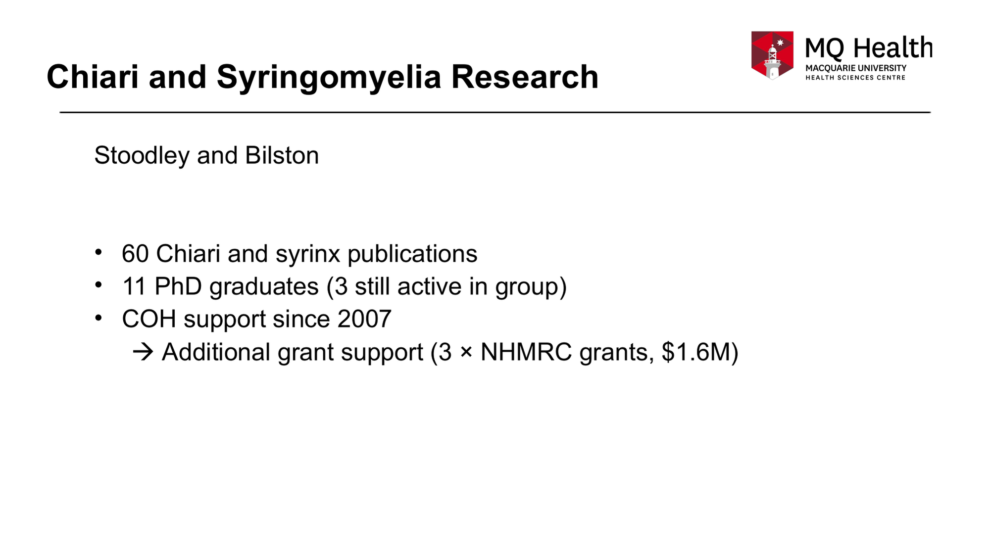
Stoodley and Bilston
60 Chiari and syrinx publications
11 PhD graduates (3 still active in group)
COH support since 2007
 Additional grant support (3 × NHMRC grants, $1.6M)
    Slide 8 — tap to see it full size

    Note the multiplying effect of the research, from increasing the scientists and surgeons interested in Chiari malformation to attracting much larger government grants to accelerate our research. Stoodley & Bilston are our two primary researchers in Sydney.
  9. Chiari and Syringomyelia Research
CHIARI
SYRINGOMYELIA
CLINICAL
1.	King V, Liu S, Russo C, Jayasekara M, Stoodley M, Di Ieva A. Use of Artificial Intelligence in the Prediction of Chiari Malformation Type 1 Recurrence After Posterior Fossa Decompressive Surgery.   Cureus. . 2024;16(5):e60879. doi:10.7759/cureus.60879
2.	Lloyd RA, Stoodley MA, Bilston LE. Statistical shape models of the posterior cranial fossa and hindbrain volumes may provide a more robust clinical metric for Chiari malformation.   J Biomech. . 2022137doi:10.1016/j.jbiomech.2022.111093
3.	Tanaka KW, Russo C, Liu S, Stoodley MA, Di Ieva A. Use of deep learning in the MRI diagnosis of Chiari malformation type I.   Neuroradiology. . 2022;64(8):1585-1592. doi:10.1007/s00234-022-02921-0
4.	Dawes BH, Lloyd RA, Rogers JM, Magnussen JS, Bilston LE, Stoodley MA. Cerebellar Tissue Strain in Chiari Malformation with Headache.   World Neurosurg. . 2019130doi:10.1016/j.wneu.2019.05.211
5.	Houston JR, Allen PA, Rogers JM, et al. Type I Chiari malformation, RBANS performance, and brain morphology: Connecting the dots on cognition and macrolevel brain structure.   Neuropsychology. . 2019;33(5):725-738. doi:10.1037/neu0000547
6.	Rogers JM, Savage G, Stoodley MA. A Systematic Review of Cognition in Chiari I Malformation.   Neuropsychol Rev. . 2018;28(2):176-187. doi:10.1007/s11065-018-9368-6
1.	Johnson L, Bartlett-Tomasetig F, Fok S, et al. A novel method to quantify perivascular space enlargement near the syrinx in a rodent model of post-traumatic syringomyelia.   Sci. rep.. . 2023;13(1):15043. doi:10.1038/s41598-023-42275-y
2.	Berliner JA, Lam MA, Najafi E, Hemley SJ, Bilston LE, Stoodley MA. Aquaporin-4 expression and modulation in a rat model of post-traumatic syringomyelia.   Sci. rep.. . 2023;13(1):9662. doi:10.1038/s41598-023-36538-x
3.	Liu S, Bilston LE, Flores Rodriguez N, et al. Changes in intrathoracic pressure, not arterial pulsations, exert the greatest effect on tracer influx in the spinal cord.   Fluids Barriers CNS. . 2022;19(1):14. doi:10.1186/s12987-022-00310-6
4.	Liu S, Bilston LE, Stoodley MA, Hemley SJ. Tachycardia and hypertension enhance tracer efflux from the spinal cord.   Fluids Barriers CNS. . 2021;18(1):47. doi:10.1186/s12987-021-00279-8
5.	Berliner J, Hemley S, Najafi E, Bilston L, Stoodley M, Lam M. Abnormalities in spinal cord ultrastructure in a rat model of post-traumatic syringomyelia.   Fluids Barriers CNS. . 2020;17(1):11. doi:10.1186/s12987-020-0171-4
6.	Lloyd RA, Stoodley MA, Fletcher DF, Bilston LE. The effects of variation in the arterial pulse waveform on perivascular flow.   J Biomech. . 201990doi:10.1016/j.jbiomech.2019.04.030
7.	Berliner JA, Woodcock T, Najafi E, et al. Effect of extradural constriction on CSF flow in rat spinal cord.   Fluids Barriers CNS. . 2019;16(1):7. doi:10.1186/s12987-019-0127-8
8.	Liu S, Lam MA, Sial A, Hemley SJ, Bilston LE, Stoodley MA. Fluid outflow in the rat spinal cord: the role of perivascular and paravascular pathways.   Fluids Barriers CNS. . 2018;15(1):13. doi:10.1186/s12987-018-0098-1
9.	Yeo J, Cheng S, Hemley S, Lee BB, Stoodley M, Bilston L. Characteristics of CSF Velocity-Time Profile in Posttraumatic Syringomyelia.   AJNR Am J Neuroradiol. . 2017;38(9):1839-1844. doi:10.3174/ajnr.A5304
10.	Najafi E, Bilston LE, Song X, et al. Longitudinal measurements of syrinx size in a rat model of posttraumatic syringomyelia.   J Neurosurg Spine. . 2016;24(6):941-8. doi:10.3171/2015.10.SPINE15538
11.	Wong JH, Song X, Hemley SJ, Bilston LE, Cheng S, Stoodley MA. Direct-trauma model of posttraumatic syringomyelia with a computer-controlled motorized spinal cord impactor.   J Neurosurg Spine. . 2016;24(5):797-805. doi:10.3171/2015.10.SPINE15742
12.	Najafi E, Stoodley MA, Bilston LE, Hemley SJ. Inwardly rectifying potassium channel 4.1 expression in post-traumatic syringomyelia.   Neuroscience. . 2016317doi:10.1016/j.neuroscience.2016.01.001
13.	Leung V, Magnussen JS, Stoodley MA, Bilston LE. Cerebellar and hindbrain motion in Chiari malformation with and without syringomyelia.   J Neurosurg Spine. . 2016;24(4):546-55. doi:10.3171/2015.8.SPINE15325
14.	Cheng S, Fletcher D, Hemley S, Stoodley M, Bilston L. Effects of fluid structure interaction in a three dimensional model of the spinal subarachnoid space.   J Biomech. . 2014;47(11):2826-30. doi:10.1016/j.jbiomech.2014.04.027
15.	Clarke EC, Fletcher DF, Stoodley MA, Bilston LE. Computational fluid dynamics modelling of cerebrospinal fluid pressure in Chiari malformation and syringomyelia.   J Biomech. . 2013;46(11):1801-9. doi:10.1016/j.jbiomech.2013.05.013
16.	Clarke EC, Stoodley MA, Bilston LE. Changes in temporal flow characteristics of CSF in Chiari malformation Type I with and without syringomyelia: implications for theory of syrinx development.   J Neurosurg. . 2013;118(5):1135-40. doi:10.3171/2013.2.JNS12759
17.	Hemley SJ, Bilston LE, Cheng S, Chan JN, Stoodley MA. Aquaporin-4 expression in post-traumatic syringomyelia.   J Neurotrauma. . 2013;30(16):1457-67. doi:10.1089/neu.2012.2614
18.	Hemley SJ, Bilston LE, Cheng S, Stoodley MA. Aquaporin-4 expression and blood-spinal cord barrier permeability in canalicular syringomyelia.   J Neurosurg Spine. . 2012;17(6):602-12. doi:10.3171/2012.9.SPINE1265
19.	Wong J, Hemley S, Jones N, Cheng S, Bilston L, Stoodley M. Fluid outflow in a large-animal model of posttraumatic syringomyelia.   Neurosurgery. . 2012;71(2):474-80; discussion 480. doi:10.1227/NEU.0b013e31825927d6
20.	Cheng S, Stoodley MA, Wong J, Hemley S, Fletcher DF, Bilston LE. The presence of arachnoiditis affects the characteristics of CSF flow in the spinal subarachnoid space: a modelling study.   J Biomech. . 2012;45(7):1186-91. doi:10.1016/j.jbiomech.2012.01.050
21.	Tu J, Liao J, Stoodley MA, Cunningham AM. Reaction of endogenous progenitor cells in a rat model of posttraumatic syringomyelia.   J Neurosurg Spine. . 2011;14(5):573-82. doi:10.3171/2011.1.SPINE09491
22.	Tu J, Liao J, Stoodley MA, Cunningham AM. Differentiation of endogenous progenitors in an animal model of post-traumatic syringomyelia.   Spine. . 2010;35(11):1116-21. doi:10.1097/BRS.0b013e3181ba6ce4
23.	Bilston LE, Stoodley MA, Fletcher DF. The influence of the relative timing of arterial and subarachnoid space pulse waves on spinal perivascular cerebrospinal fluid flow as a possible factor in syrinx development.   J Neurosurg. . 2010;112(4):808-13. doi:10.3171/2009.5.JNS08945
24.	Hemley SJ, Tu J, Stoodley MA. Role of the blood-spinal cord barrier in posttraumatic syringomyelia.   J Neurosurg Spine. . 2009;11(6):696-704. doi:10.3171/2009.6.SPINE08564
25.	Bertram CD, Bilston LE, Stoodley MA. Tensile radial stress in the spinal cord related to arachnoiditis or tethering: a numerical model.   Med Biol Eng Comput. . 2008;46(7):701-7. doi:10.1007/s11517-008-0332-0
26.	Brodbelt A, Stoodley M. CSF pathways: a review.   Br J Neurosurg. . 2007;21(5):510-20. doi:10.1080/02688690701447420
27.	Bilston LE, Fletcher DF, Stoodley MA. Focal spinal arachnoiditis increases subarachnoid space pressure: a computational study.   Clin Biomech. . 2006;21(6):579-84. doi:10.1016/j.clinbiomech.2006.01.009
28.	Bertram CD, Brodbelt AR, Stoodley MA. The origins of syringomyelia: numerical models of fluid/structure interactions in the spinal cord.   J Biomech Eng. . 2005;127(7):1099-109. doi:10.1115/1.2073607
29.	Brodbelt AR, Stoodley MA, Watling A, et al. The role of excitotoxic injury in post-traumatic syringomyelia.   J Neurotrauma. . 2003;20(9):883-93. doi:10.1089/089771503322385818
30.	Brodbelt AR, Stoodley MA, Watling AM, Tu J, Burke S, Jones NR. Altered subarachnoid space compliance and fluid flow in an animal model of posttraumatic syringomyelia.   Spine. . 2003;28(20):E413-9. doi:10.1097/01.BRS.0000092346.83686.B9
31.	Bilston LE, Fletcher DF, Brodbelt AR, Stoodley MA. Arterial pulsation-driven cerebrospinal fluid flow in the perivascular space: a computational model.   Comput Methods Biomech Biomed Engin. . 2003;6(4):235-41. doi:10.1080/10255840310001606116
32.	Brodbelt AR, Stoodley MA, Watling AM, Tu J, Jones NR. Fluid flow in an animal model of post-traumatic syringomyelia.   Eur Spine J. . 2003;12(3):300-6. doi:10.1007/s00586-002-0492-9
33.	Yang L, Jones NR, Stoodley MA, Blumbergs PC, Brown CJ. Excitotoxic model of post-traumatic syringomyelia in the rat.   Spine. . 2001;26(17):1842-9. doi:10.1097/00007632-200109010-00004
34.	Stoodley MA, Jones NR, Yang L, Brown CJ. Mechanisms underlying the formation and enlargement of noncommunicating syringomyelia: experimental studies.   Neurosurg. focus. . 2000;8(3):E2. doi:10.3171/foc.2000.8.3.2
35.	Stoodley MA. Pathophysiology of syringomyelia.   J Neurosurg. . 2000;92(6):1069-70; author reply 1071-3. Cited in: Ovid MEDLINE(R) at http://ovidsp.ovid.com/ovidweb.cgi?T=JS&PAGE=reference&D=med4&NEWS=N&AN=10839277. Accessed July 08, 2025.
36.	Stoodley MA, Gutschmidt B, Jones NR. Cerebrospinal fluid flow in an animal model of noncommunicating syringomyelia.   Neurosurgery. . 1999;44(5):1065-75; discussion 1075-6. doi:10.1097/00006123-199905000-00068
37.	Storer KP, Toh J, Stoodley MA, Jones NR. The central canal of the human spinal cord: a computerised 3-D study.   J Anat. . 1998192 ( Pt 4)doi:10.1046/j.1469-7580.1998.19240565.x
38.	Stoodley MA, Jones NR, Brown CJ. Evidence for rapid fluid flow from the subarachnoid space into the spinal cord central canal in the rat.   Brain Res. . 1996;707(2):155-64. doi:10.1016/0006-8993(95)01228-1
1.	Park RJ, Unnikrishnan S, Berliner J, Magnussen J, Liu S, Stoodley MA. Cerebellar Tonsillar Descent Mimicking Chiari Malformation.   J. Clin. Med.. . 2023;12(8). doi:10.3390/jcm12082786
2.	Ciaramitaro P, Massimi L, Bertuccio A, et al. Diagnosis and treatment of Chiari malformation and syringomyelia in adults: international consensus document.   Neurol Sci. . 2022;43(2):1327-1342. doi:10.1007/s10072-021-05347-3
3.	Massimi L, Peretta P, Erbetta A, et al. Diagnosis and treatment of Chiari malformation type 1 in children: the International Consensus Document.   Neurol Sci. . 2022;43(2):1311-1326. doi:10.1007/s10072-021-05317-9
4.	Jeffree RL, Stoodley MA. Management of Chiari in pregnancy.   J Clin Neurosci. . 202183doi:10.1016/j.jocn.2020.10.025
5.	Davidson KA, Rogers JM, Stoodley MA. Syrinx to Subarachnoid Shunting for Syringomyelia.   World Neurosurg. . 2018110doi:10.1016/j.wneu.2017.09.205
6.	Davidoff CL, Liu S, Wong JHY, Koustais S, Rogers JM, Stoodley MA. Treatment of Syringomyelia in Patients with Arachnoiditis at the Craniocervical Junction.   World Neurosurg. . 2017107doi:10.1016/j.wneu.2017.08.064
7.	Li AE, Wilkinson MD, McGrillen KM, Stoodley MA, Magnussen JS. Clinical Applications of Cine Balanced Steady-State Free Precession MRI for the Evaluation of the Subarachnoid Spaces.   Clin Neuroradiol. . 2015;25(4):349-60. doi:10.1007/s00062-015-0383-1
8.	Brodbelt AR, Stoodley MA. Syringomyelia and the arachnoid web.   Acta Neurochir (Wien). . 2003;145(8):707-11; discussion 711. doi:10.1007/s00701-003-0071-9
9.	Brodbelt AR, Stoodley MA. Post-traumatic syringomyelia: a review.   J Clin Neurosci. . 2003;10(4):401-8. doi:10.1016/s0967-5868(02)00326-0
    Slide 9 — tap to see it full size

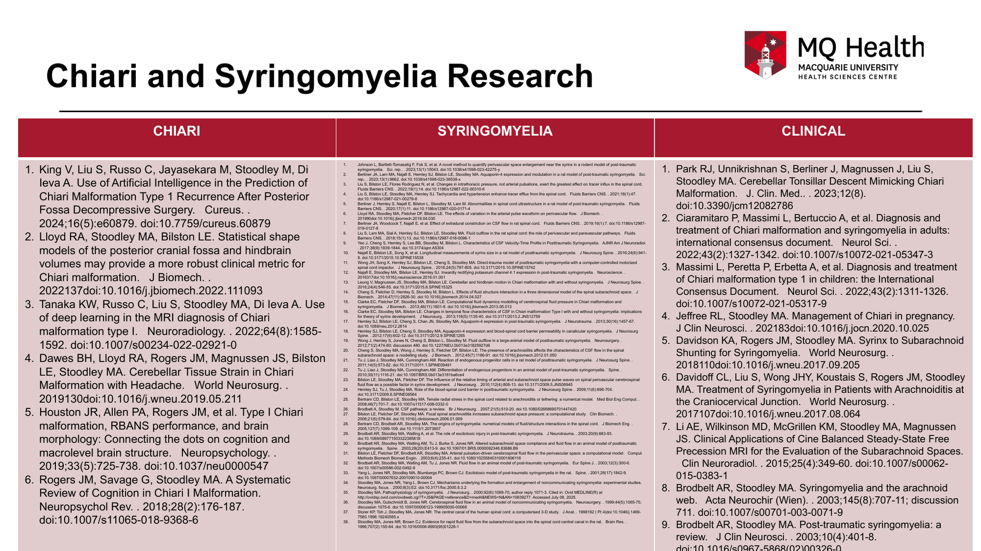
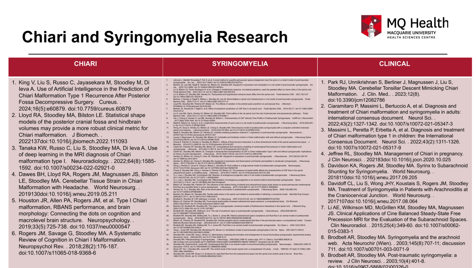
    The number of studies and the quality of the publications speak for themselves. This research has taken us from “Unknown Pathophysiology” and “No Clear Treatment Goals” to consistent successful surgeries at one centre. Out next step is to determine the technical outcomes of surgery to share with neurosurgeons worldwide!!!

    Column of Hope funded peer-reviewed research studies, with a link to each paper
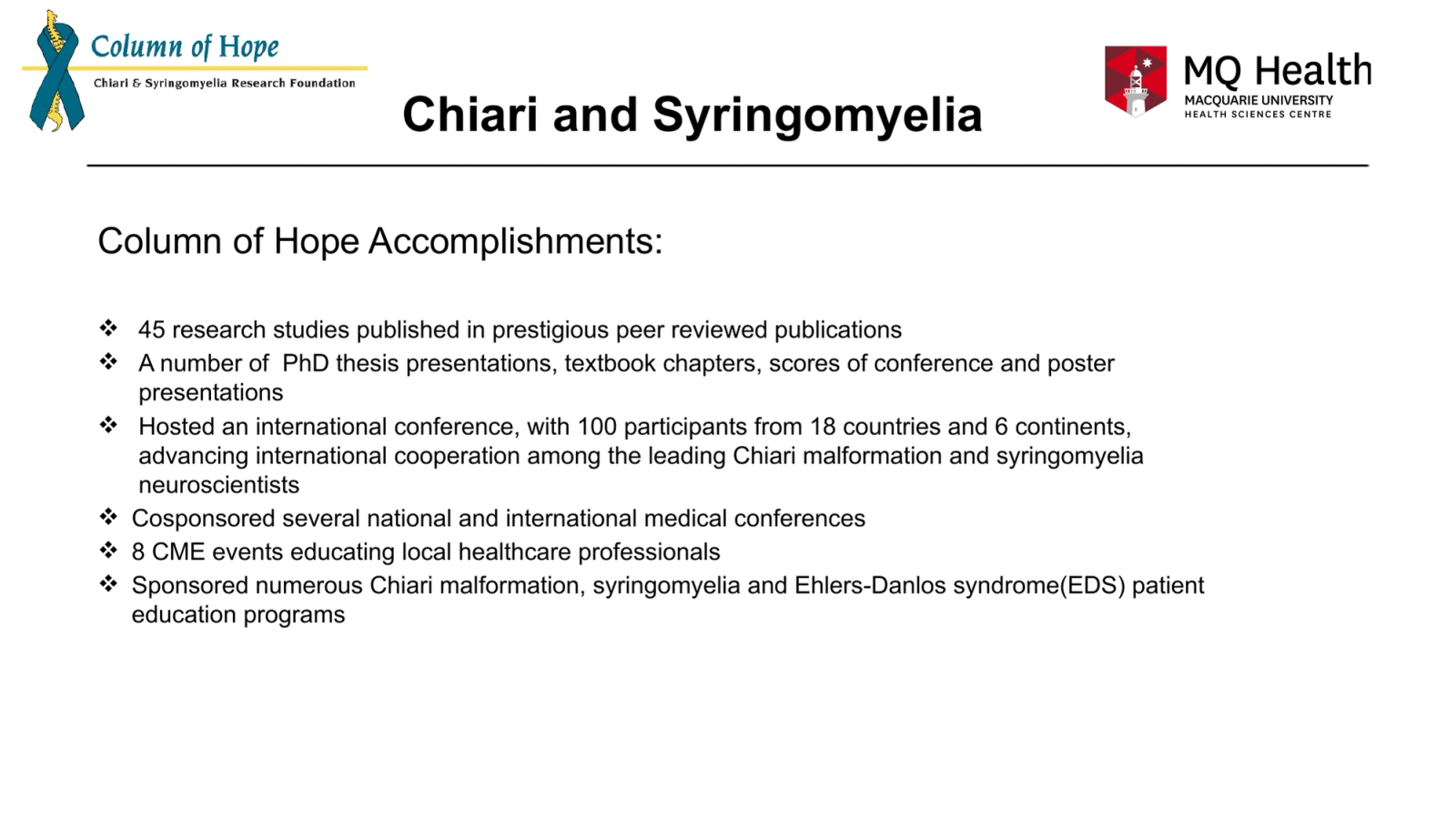
  10. Chiari and Syringomyelia
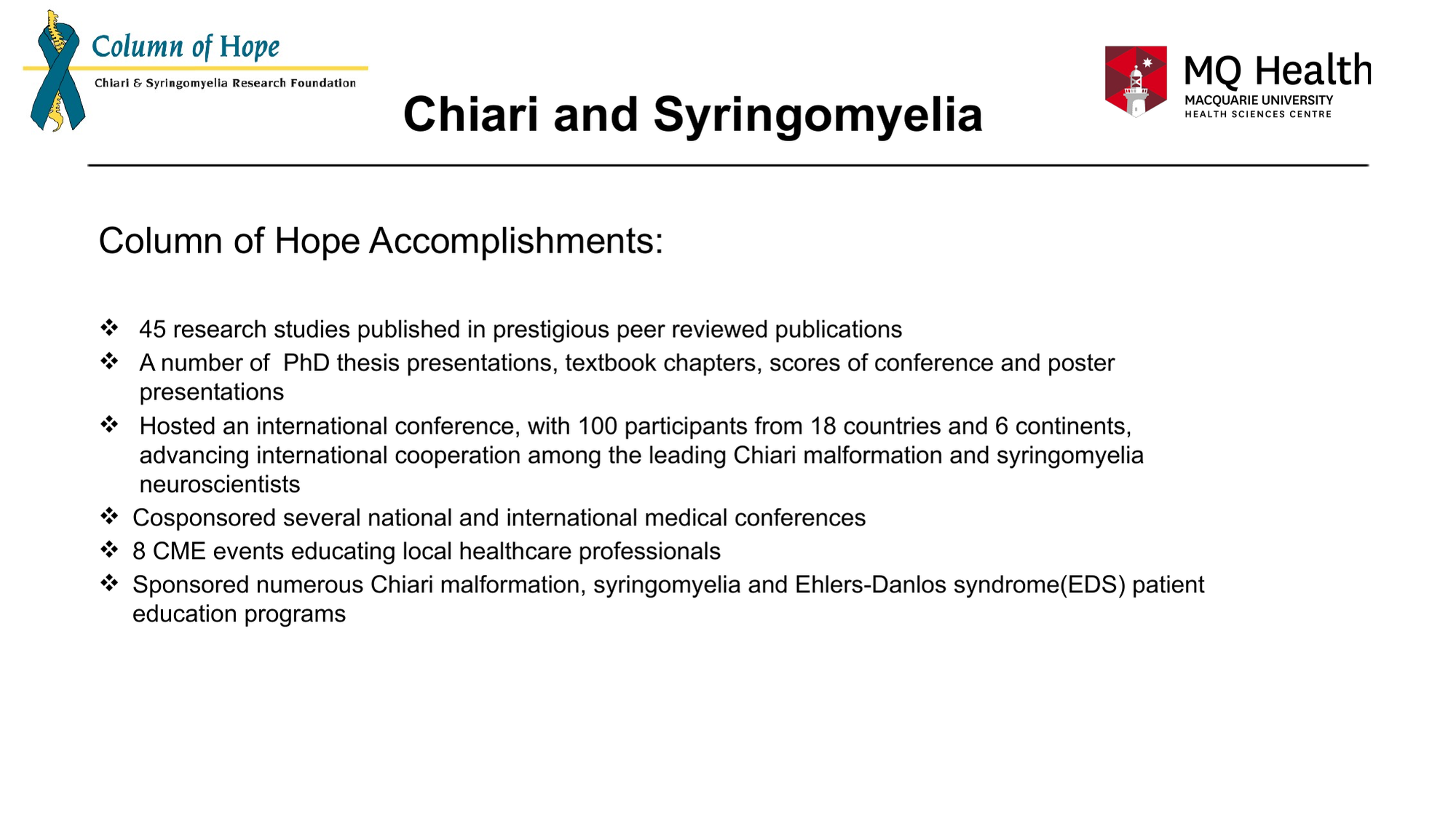
Column of Hope Accomplishments:
45 research studies published in prestigious peer reviewed publications
A number of  PhD thesis presentations, textbook chapters, scores of conference and poster presentations
Hosted an international conference, with 100 participants from 18 countries and 6 continents, advancing international cooperation among the leading Chiari malformation and syringomyelia neuroscientists
Cosponsored several national and international medical conferences
8 CME events educating local healthcare professionals
Sponsored numerous Chiari malformation, syringomyelia and Ehlers-Danlos syndrome(EDS) patient education programs
    Slide 10 — tap to see it full size

    Column of Hope is eternally grateful to its donors for making this possible!

  11. Slide 11
    Slide 11 — tap to see it full size

    Our Team in Sydney Australia. Seeds from Column of Hope have grown into a deep-rooted team!!!
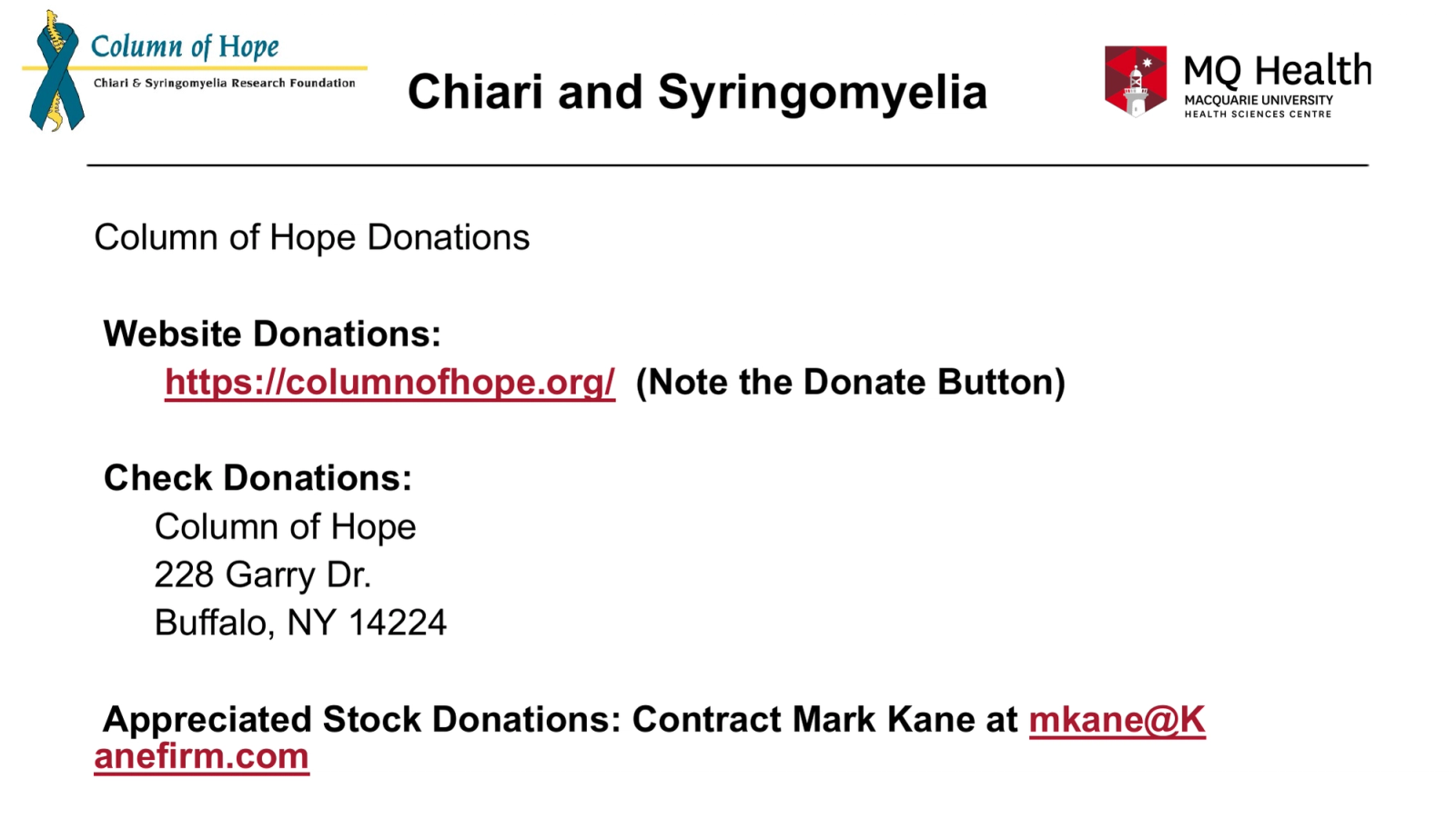
  12. Chiari and Syringomyelia
Column of Hope Donations
Website Donations:
https://columnofhope.org/  (Note the Donate Button)
Check Donations:
Column of Hope
228 Garry Dr.
Buffalo, NY 14224
Appreciated Stock Donations: Contract Mark Kane at mkane@Kanefirm.com
    Slide 12 — tap to see it full size

    At Column of Hope, we feel we have come much farther in the last 20 years than all the progress made combined in the nearly 460 years before that. We now need to run well in the anchor leg of this relay race to allow patients in the US and around the world to receive the benefits of the progress we have made in the lab and one centre! Your support for this anchor leg will be greatly appreciated.
Take it with you

Candid transparency sealDonate